File:M aqaa029i0001.jpeg
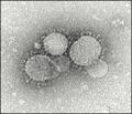
Electron microscope image of coronavirus [1].
Description
In the past two decades, the world has seen three coronaviruses emerge and cause outbreaks that have caused considerable global health consternation. Coronaviruses are enveloped, nonsegmented, single-stranded, positive-sense RNA viruses that have a characteristic appearance on electron microscopy negative staining Image 1. As a matter of fact, the characteristic electron microscopy appearance was the clue to amplify and sequence nucleic acids from Dr Urbani’s (one of the health care providers who died of severe acute respiratory syndrome [SARS] in 2003) respiratory specimen using a consensus coronavirus primer.1 The sequence of the virus was significantly different from other coronaviruses known to cause human disease at the time. The virus was ultimately named SARS-CoV, as febrile patients had severe acute respiratory syndrome and could present with pneumonia and lower respiratory symptoms such as cough and dyspnea.2 The SARS-CoV outbreak started in Guangdong, China, and spread to many countries in Southeast Asia, North America, Europe, and South Africa. Transmission was primarily person to person through droplets that occurred during coughing or sneezing, through personal contact (shaking hands), or by touching contaminated surfaces. Of note, health professionals were particularly at risk of acquiring the disease, as transmission also occurred if isolation precautions were not followed and during certain procedures. The last case of SARS-CoV occurred in September 2003, after having infected over 8,000 persons and causing 774 deaths with a case fatality rate calculated at 9.5%.
References
- ↑ https://academic.oup.com/ajcp/article/153/4/420/5735509 Three Emerging Coronaviruses in Two Decades: The Story of SARS, MERS, and Now COVID-19 Jeannette Guarner, MD. American Journal of Clinical Pathology, Volume 153, Issue 4, April 2020, Pages 420–421, https://doi.org/10.1093/ajcp/aqaa029 Published: 13 February 2020 In the past two decades, the world has seen three coronaviruses emerge and cause outbreaks that have caused considerable global health consternation. Coronaviruses are enveloped, nonsegmented, single-stranded, positive-sense RNA viruses that have a characteristic appearance on electron microscopy negative staining Image 1. As a matter of fact, the characteristic electron microscopy appearance was the clue to amplify and sequence nucleic acids from Dr Urbani’s (one of the health care providers who died of severe acute respiratory syndrome [SARS] in 2003) respiratory specimen using a consensus coronavirus primer.1 The sequence of the virus was significantly different from other coronaviruses known to cause human disease at the time. The virus was ultimately named SARS-CoV, as febrile patients had severe acute respiratory syndrome and could present with pneumonia and lower respiratory symptoms such as cough and dyspnea.2 The SARS-CoV outbreak started in Guangdong, China, and spread to many countries in Southeast Asia, North America, Europe, and South Africa. Transmission was primarily person to person through droplets that occurred during coughing or sneezing, through personal contact (shaking hands), or by touching contaminated surfaces. Of note, health professionals were particularly at risk of acquiring the disease, as transmission also occurred if isolation precautions were not followed and during certain procedures. The last case of SARS-CoV occurred in September 2003, after having infected over 8,000 persons and causing 774 deaths with a case fatality rate calculated at 9.5%.
File history
Click on a date/time to view the file as it appeared at that time.
| Date/Time | Thumbnail | Dimensions | User | Comment | |
|---|---|---|---|---|---|
| current | 16:36, 28 March 2020 |  | 520 × 448 (39 KB) | T (talk | contribs) |
- You cannot overwrite this file.
File usage
There are no pages that link to this file.
