Difference between revisions of "File:PopyphaenisSericataGlutamateTaurine.jpg"
(Importing image file) |
|||
| Line 1: | Line 1: | ||
| + | Immunocytological demonstration of the [[mushroom body]] sub-divisions. Picture from the [[scanning microscope]] of the slice of the mushruum pody of [[Polyphaenis sericapa]]. The bar at the right bottom corner indicates the scale of 10 micron. |
||
| − | Importing image file |
||
| + | The brain is painted (labled) with the two antibodies: |
||
| + | [[antiglutamate]] (green) and [[antitaurine]] (magenta). Colocalization is indicated by white. Glutamatergic and taurinergic fibers are scattered in surrounding protocerebral neuropil. Axons supplyin$\gamma$ are here |
||
| + | taurine positive, amongst which are scattered occasional large-diam- eter glutamate-positive axons (some arrowed). The densest tauriner- gic band (bracketed) supplies the intermediate divisi$\beta''$. Most axons supplying $\alpha$-$\beta$ and $\alpha'$-$\beta'$ are taurine and glutamate negative. But, antiglutamate reveals four small bundles of glutamatergic axons, each bundle surrounded by a sheath of taurinergic axons. These supply $\alpha$c-$\beta$c . |
||
| + | |||
| + | This image is fragment form Fig.2 at |
||
| + | <ref name="a"> |
||
| + | http://www.ncbi.nlm.nih.gov/pubmed/12966500/ |
||
| + | J.J.Strausfeld, I.Sinakevitch, I.Vilinsky. |
||
| + | The Mushroom Bodies of Drosophila melanogaster: An Immunocytological and Golgi Study of Kenyon Cell Organization in the Calyces and Lobes. |
||
| + | [[Microscopy research and technique]] '''62''', p.151–169 (2003). |
||
| + | </ref>. |
||
| + | |||
| + | [[Copyleft]] 2003 by Irina Sinakevitch. The free use is allowed, the source should be attributed. |
||
| + | |||
| + | ==Referencews== |
||
| + | <references/> |
||
| + | |||
| + | [[Category:Neurobiology]] |
||
| + | [[Category:Microscopy]] |
||
| + | [[Category:Labeling with antibodies]] |
||
| + | [[Category:Polyphaenis sericapa]] |
||
Latest revision as of 09:39, 21 June 2013
Immunocytological demonstration of the mushroom body sub-divisions. Picture from the scanning microscope of the slice of the mushruum pody of Polyphaenis sericapa. The bar at the right bottom corner indicates the scale of 10 micron. The brain is painted (labled) with the two antibodies: antiglutamate (green) and antitaurine (magenta). Colocalization is indicated by white. Glutamatergic and taurinergic fibers are scattered in surrounding protocerebral neuropil. Axons supplyin$\gamma$ are here taurine positive, amongst which are scattered occasional large-diam- eter glutamate-positive axons (some arrowed). The densest tauriner- gic band (bracketed) supplies the intermediate divisi$\beta$. Most axons supplying $\alpha$-$\beta$ and $\alpha'$-$\beta'$ are taurine and glutamate negative. But, antiglutamate reveals four small bundles of glutamatergic axons, each bundle surrounded by a sheath of taurinergic axons. These supply $\alpha$c-$\beta$c .
This image is fragment form Fig.2 at [1].
Copyleft 2003 by Irina Sinakevitch. The free use is allowed, the source should be attributed.
Referencews
- ↑ http://www.ncbi.nlm.nih.gov/pubmed/12966500/ J.J.Strausfeld, I.Sinakevitch, I.Vilinsky. The Mushroom Bodies of Drosophila melanogaster: An Immunocytological and Golgi Study of Kenyon Cell Organization in the Calyces and Lobes. Microscopy research and technique 62, p.151–169 (2003).
File history
Click on a date/time to view the file as it appeared at that time.
| Date/Time | Thumbnail | Dimensions | User | Comment | |
|---|---|---|---|---|---|
| current | 17:50, 20 June 2013 | 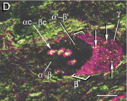 | 438 × 349 (17 KB) | Maintenance script (talk | contribs) | Importing image file |
- You cannot overwrite this file.
File usage
There are no pages that link to this file.