Search results
Create the page "Microscopy" on this wiki! See also the search results found.
- ...n are related with use of [[confocal optics]] and/or the [[interferometric microscopy]] Y.Kuznetsova; A.Neumann, S.R.Brueck (2007). "Imaging interferometric microscopy–approaching the linear systems limits of optical resolution". [[Optics Ex12 KB (1,898 words) - 18:25, 30 July 2019
- ...[Yulya function]] in microscopy and applicability of the [[interferometric microscopy]]. However, this option was not mentioned in the original slideshow by prof5 KB (698 words) - 15:01, 20 June 2013
- ...ccupied (for the dependence, that has application in the [[interferometric microscopy]]), so, the superfunction should have another name. [[Shoko]] agreed that h10 KB (1,479 words) - 05:27, 16 December 2019
- ...en occupied (denoting the dependence that appears in the [[interferometric microscopy]]), the function (1) is named after Shoko (with her permission).10 KB (1,507 words) - 18:25, 30 July 2019
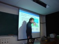
File:DSC01577henryk1.jpg ...12 Sugadaira summer school]]. Henryk san about the nonlinear spectroscopic microscopy.(3,648 × 2,736 (1.76 MB)) - 09:42, 21 June 2013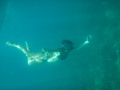
File:KuznetsovaP1205981.JPG ...Kuznetsova]] at Thailand during the unofficial workshop on the holographic microscopy, 2012.(2,560 × 1,920 (986 KB)) - 09:40, 21 June 2013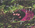
File:PopyphaenisSericataGlutamateTaurine.jpg [[Microscopy research and technique]] '''62''', p.151–169 (2003). [[Category:Microscopy]](438 × 349 (17 KB)) - 09:39, 21 June 2013- ...ristic appearance of virions (the infective form of the virus) by electron microscopy, which have a fringe of large, bulbous surface projections creating an imag38 KB (4,474 words) - 18:01, 2 April 2020
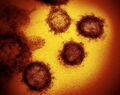
File:SARS-CoV with coronaFragment.jpg [[Electron microscopy]] [[Category:Electron microscopy]](1,431 × 1,133 (178 KB)) - 20:52, 24 February 2020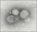
File:M aqaa029i0001.jpeg ...egative staining Image 1. As a matter of fact, the characteristic electron microscopy appearance was the clue to amplify and sequence nucleic acids from Dr Urban ...egative staining Image 1. As a matter of fact, the characteristic electron microscopy appearance was the clue to amplify and sequence nucleic acids from Dr Urban(520 × 448 (39 KB)) - 16:38, 28 March 2020


